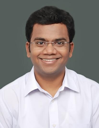
Faculty Profile

Gokulakrishnan Periakaruppan

Professor

Qualification

MBBS | M.D

Contact Info

[email protected]

Bio

Curriculum Vitae


Personal details and contact information
Name Gokulakrishnan Periakaruppan
Subspeciality interests Whole body oncoimaging including PET-CT, non-vascular interventions, acute body imaging and general radiology.


Registrations
GMC number 7906453

Specialist Registered Radiologist in UK

Tamil Nadu State Medical Council – India 77996


Memberships
The Royal College of Radiologists
Indian Radiological and Imaging Association
National University of Singapore
Radiological Society of North America
Society of Oncologic Imaging in India
European Society of Oncologic Imaging
European Society of Radiology


Qualifications
M.B.B.S (India)
M.D. Radiodiagnosis (India)
Fellow of Royal College of Radiologists (London)
M. Med. Radiology (Singapore)


Employment history
1/9/2025 to present date –              Senior Consultant Radiologist and Professor of Radiology, SRIHER.
11/08/2023 – 31/07/2025 Title: Consultant Radiologist (Full-time fixed term post) Location: Royal Preston Hospital, Lancashire Teaching Hospitals NHS Foundation Trust, UK.

Supervisor: Dr Gagan Thind Job description:

·         Independent reporting as a consultant radiologist working primarily with special interest in

oncoimaging and gastrointestinal radiology.

·         Participation in weekly hematology MDT.

·         Participation in on-call duties.

·         Performing image guided interventions.


·         Training of specialty trainees in radiology.
1/4/2021-31/07/2023 Title: Professor and consultant Radiologist

Location: Sri Ramachandra Institute for higher education and research

Supervisor: Prof. Rajeswaran. R Job description:

·         Rotations through ultrasound, CT, MRI, PET/CT modalities – unsupervised and independent reporting.

·         Attending on-call duties and reporting emergency cases.

·         Teaching and training radiology post graduates.

·         Teaching and training of M.B.B.S students.

·         Participation in weekly oncology MDT and conducting monthly radiology-general medicine and radiology- endocrine monthly meetings.

·         Independent performing image guided interventions.

·         Participation in management activities like rota co- ordinator for staff, medical staff credential committee.

·         Internal and external examiner for radiology post graduate trainees for final exam.

1/4/2016- 31/03/2021 Title: Associate professor and consultant radiologist. Location: Sri Ramachandra Institute for higher education and research.

Supervisor: Prof. P.M. Venkata Sai Job description:

·         Independent and unsupervised radiology reporting of all modalities.

·         Attending on-call duties and reporting emergency cases.

·         Monitoring of image protocols and oversight of radiology technicians.

·         Supervise the work of radiology trainees and junior faculty.

·         Moderate and conduct classes for radiology post graduates, B.Sc. and M.Sc. Medical Imaging Technology students.

·         Performing image guided interventions without supervision.

·         Examiner for radiology residents and allied health science students.

·         Participation in weekly oncology MDT and conducting radiology-general medicine monthly meetings.

16/6/2010- 31/03/2016 Title: Assistant professor and consultant radiologist. Location: Sri Ramachandra Medical College and Hospital Supervisor: Prof. P.M. Venkata Sai

Job description:

·         Independent radiology reporting covering all different modalities.


·         Attending on-call duties and reporting emergency duties.

·         Teaching of radiology interns and medical imaging technology students.

·         Performing image guided interventions independently.

March 2007 – April 2010 Title: Junior Resident in Radiology

Location: Sri Ramachandra Medical College and Hospital, Porur, Chennai, Tamil Nadu, India.

Supervisor: Prof. Santosh Joseph Job description:

Completed rotations in

·         Radiography (positioning and techniques,

fluoroscopic procedures including IVP, MCU, AUG,

barium studies, sinogram, fistulograms, cystograms, opposing urethrograms, sialograms and NJ tube insertion)

·         Mammography

·         Ultrasound

·         Computed Tomography

·         Magnetic Resonance Imaging

·         Nuclear Medicine

·         Neurointerventional radiology


Assisted and performed image guided drainages, FNACs, biopsies under supervision of consultants and independently.

Reported and performed ultrasound and Dopplers under consultant supervision and independently.

Attended on-call duties and reporting emergency cases.

23.02.2006 Primary Medical Qualification (M.B.B.S) awarded.
28.12.2005 M.B.B.S passed date


Gap in employment
February 2006 – February 2007 Gap while preparing for post graduate national entrance examination.
25/04/2010 – 15/06/2010 Gap while applying for employment post training.

No gap that spans more than 28 days after securing a job since

16-6-2010.


Research experience
Thesis guide post graduate students in following research topics:

·         Comparison of diagnostic yield between CT guided and PET/CT image guided biopsies.

·         Role of sheer wave elastography in predicting indeterminate nodules by comparing with fine

needle aspiration cytology.


·         Analysis of pneumothorax incidence and its risk factors after transthoracic biopsy of lung lesions under CT guidance.

·         Shape and volumetric analysis of pituitary gland on MRI in paediatric age group in Indian population.

·         Analysis of correlation between SUV max and staging of tumour in patients with carcinoma stomach.


Publications
Goyal, Nishita & Periakaruppan, Gokulakrishnan & Murali, Arunan & Dennis, Leena & Rangasami, Rajeswaran & Dev, Bhawna. (2023). Association of Standardized Uptake Values of Primary Breast Cancer on [18F] FDG PET/CT With

Immunohistochemistry and Molecular Subtypes: Correlation of SUV of primary BC with molecular subtypes. Archives of Breast Cancer. 175-186. 10.32768/abc.2023102175-186.

Rajakumar Sibhithran & Murali Arunan & Periakaruppan

Gokulakrishnan & Rangasami Rajeswaran (2023). Pulmonary hot- clot artifacts mimicking as metastatic lung lesions in 18F- fluorodeoxyglucose positron emission tomography/computed tomography – A case series. Case Reports in Clinical Radiology. 1. 5-9. 10.25259/CRCR_36_2022.

Mannam, Pallavi & Murali, Arunan & Periakaruppan,

Gokulakrishnan & Venkatachalapathy, Easwaramoorthy & Sai,

PulivadulaMohanarangam. (2022). Radiomic Analysis of Positron- Emission Tomography and Computed Tomography Images to

Differentiate between Multiple Myeloma and Skeletal

Metastases. Indian Journal of Nuclear Medicine. 37. 217. 10.4103/ijnm.ijnm_111_21.

Chidurala R, Sneha LM, Kothandan BT, Arunachalam R,

Jeyabalakrishnan S, Arumugam VG, et al. Inflammatory myofibroblastic tumour of an unusual site in an infant. Int J Contemp Pediatr 2022; 9:1208-11.

John C, Venkatasai J, Kondaveeti SS, Murali A, Periakaruppan G, E V, Meenakshisundaram M, Ambalathandi RC, Masilamani H.

Prognostic significance of metabolic tumour volume and

maximum standard uptake value of fluor-18-fluorodeoxyglucose positron emission tomography with computed tomography in nasopharyngeal carcinoma. Contemp Oncol (Pozn).

2021;25(3):153-159. doi: 10.5114/wo.2021.109620. Epub 2021

Oct 1. PMID: 34729034; PMCID: PMC8547178.

Tewari S, Chinnappan S, Gokulakrishnan PR, Gupta C. Radiologic exploration of the morphometric and morphological features of

the carotid canal. J Taibah Univ Med Sci. 2022 May 13;17(6):936-942. doi: 10.1016/j.jtumed.2022.04.013. PMID: 36212579; PMCID: PMC9519627.


Pradeep, Nambron & Ayub, Irfan & Krishnaswamy, Madhavan & Periakaruppan, Gokulakrishnan. (2021). Bilevel positive airway pressure in tracheobronchomalacia. BMJ case reports. 14.

10.1136/bcr-2021-246331.

Pallavi M, Murali A, Periakaruppan G, Easwaramoorthy V, Mohanarangam VSP. Correlation between Reference Tissue Normalized 18F-Fluorodeoxyglucose Positron Emission

Tomography/Computed Tomography Standardized Uptake Value Max of Nodal and Extranodal Sites in Lymphomas: An Empirical Study. Indian J Nucl Med. 2021 Oct-Dec;36(4):371-376. doi:

10.4103/ijnm.ijnm_74_21. Epub 2021 Dec 15. PMID: 35125754; PMCID: PMC8771065.

Basetti, B., Periakaruppan, G., Murali, A. et al. Breast involvement in granulomatosis with polyangiitis: a case report. Egypt J Radiol Nucl Med 52, 193 (2021). https://doi.org/10.1186/s43055-021-00569-z
Pabla, Harleen & Periakaruppan, Gokulakrishnan & Murali, Arunan & P.M., Venkata. (2021). Role of PET-CT in Aiding Diagnosis of Various Neurological Conditions – A Case Series.

Journal of Evolution of Medical and Dental Sciences. 10. 440-446. 10.14260/jemds/2021/97.

Periakaruppan, Gokulakrishnan, et al. “Clinical Correlation of Maximum Standardised Uptake Value (SUVmax) in Positron Emission Tomography–Computed Tomography (PET-CT) with

Initial Disease Staging of Carcinoma Breast–An Observational Study at Indian Tertiary Care Centre.” Journal of Evolution of Medical and Dental Sciences, vol. 9, no. 17, 27 Apr. 2020.

Jeyarani, Dr & Sneha, Dr & Jayaraman, Dhaarani & Gunabooshanam, Barathi & Periakaruppan, Gokulakrishnan & Kumaravel, Dr. (2019). Rosai–Dorfman disease presenting in a child as mediastinal lymphadenopathy. Pediatric Review:

International Journal of Pediatric Research. 6. 625-628. 10.17511/ijpr. 2019.i12.07.

Periakaruppan, Gokulakrishnan & Murali, Arunan & Raj, Bhasker & venkata sai, Pm. (2018). Correlation and Predictive Value of the FDG uptake within Primary Mass in Non-small Cell Lung Carcinoma and Incidence of Distal Metastases. International

Journal of Contemporary Medicine, Surgery and Radiology. 3. 10.21276/ijcmsr.2018.3.3.24.

Murali A, Periakaruppan G, Raj B, et al. Correlation between the FDG uptake of primary tumour and metastatic mediastinal lymphnodes in non-small cell lung carcinoma. J. Evid. Based Med. Healthc. 2018; 5(34), 2459-2462. DOI:

10.18410/jebmh/2018/507


Murali, Arunan & Periakaruppan, Gokulakrishnan & Raj, Bhasker & venkata sai, Pm. (2018). Correlation between the FDG uptake of primary tumour and metastatic mediastinal lymph nodes in non-small cell lung carcinoma. Journal of Evidence Based

Medicine and Healthcare. 5. 10.18410/jebmh/2018/507.

Periakaruppan G, Seshadri KG, Vignesh Krishna GM, Mandava R, Sai VPM, Rajendiran S. Correlation between Ultrasound-based

TIRADS and Bethesda System for Reporting Thyroid-

cytopathology: 2-year Experience at a Tertiary Care Center in India. Indian J Endocrinol Metab. 2018 Sep-Oct;22(5):651-655. doi: 10.4103/ijem.IJEM_27_18. PMID: 30294576; PMCID:

PMC6166562.

Gokulakrishnan Periakaruppan, Joe Joseph, P M Venkata Sai, Arun B Chittaranjan. Comparative study between various growth pattern of masseter muscles using ultrasound imaging system.

Medpulse international medical journal – Feb 2015. Volume 2;

issue2.

Joe Joseph, Gokulakrishnan Periakaruppan, Arun B Chitharanjan,

PM Venkata Sai. Assessment of thickness of the temporalis muscle in orthodontic patients with various growth patterns

using ultrasound imaging system. Internation journal of Recent Trends in Science and Technology; March 2015, volume 14,

issue2.

Chandramohan V, Nagarajan VP, Sathyamoorthi MS, Kumar S,

Shanmugasundaram C, Periakaruppan G, Scott JX. Posterior

reversible encephalopathy syndrome in a child with autoimmune lymphoproliferative syndrome: Case report and review of

literature. J Pediatr Neurosci. 2012 Sep;7(3):221-4. doi: 10.4103/1817-1745.106486. PMID: 23560015; PMCID: PMC3611917.


Presentations
National ·         Case discussion – chest imaging – Ramachandra Advanced Radiology Education 2019

·         MSK imaging – RARE workshop – 2018

·         Bone tumours – Ramachandra Advanced Radiology Education 2018.

·         Interesting chest x-rays- Indian Medical Association – 2017.

·         Temporal bone imaging – ASCENT 2017.

·         Case discussion in MSK radiology – Ramachandra Advanced Radiology Education 2017.

·         Imaging in gynaecology benign conditions – IRIA 2016.

·         GIT imaging – Indian Radiology Imaging Association 2016.

·         PG symposium on Wilm’s tumour – 2015.

·         PET-CT in gynaecology malignancies- institute of

reproductive medicine and women’s health – 2015.

·         Chest imaging – Ramachandra Advanced Radiology Education 2015.

·         GUT imaging – Ramachandra Advanced Radiology Education 2014.

·         Target conference – Endocrine imaging – Imaging in hypoparathyroidism – 2013.


Continuing Professional

Development (CPD)

·         Monthly discrepancy meetings -Mike Dobson’s education

·         Attending speciality board meetings

·         Conferences and courses attended as shown below.

·         RCR online CPD courses as shown in appraisals

·         Satisfactory completion of trust mandatory training programmes as shown in appraisals

·         Journal reading and updating knowledge

·         Attending monthly audits

·         Undertaken audit, reaudit and QIP.

·         Reflections from colleagues, patients and students feedbacks.


Conferences attended
International ·         Training in WHO methodology for interpretation of Pediatric chest radiographs – Agra – organised by John Hopkins university.

·         European society of oncologic imaging conference in collaboration with Indian society of oncologic imaging – 2018.

·         Radiological Society of North America, Chicago, USA – 2010.

·         Interactive CME programme by Darlington education trust, UK – 2009.


National ·         Annual national conference of Indian Radiology Imaging Association 2023

·         Annual conference of Indian Radiology Imaging Association – 2022

·         Indian Society of Gastroenterology and Abdominal Radiology – 2017

·         Advanced oncoimaging conference – 2016

·         Annual conference of Indian Radiology Imaging Association – 2016

·         CT update, Goa, India – 2015

·         Radiology simplified – 2013

·         Columbia Asia FRCR course – 2012.

·         Stanley radiology FRCR course – 2012

·         CME on MSK imaging 2010

·         ISNR, 2009

·         BRACE 2009, Chennai, India.

·         Post graduate refresher course, CMC Vellore – 2009

·         Ramachandra Advanced Radiology Education, Chennai, India -2009

·         CME on radiological interventions in gynaecology – 2008

·         CME on obstetrics ultrasound -2008

·         BRACE 2008, Chennai, India.

·         Plain X-ray interpretation course – Saveetha University 2008

·         Annual national conference of Indian Radiology Imaging Association, Bangalore, India – 2007.

·         Indian Society of Neuroradiology 2007, Mumbai, India.

·         BRACE 2007, Chennai, India.


Conferences chaired
National 75th Annual conference of Indian Radiology Imaging Association, Chennai, India -2022


Courses conducted
National Equinox, SRMC, Chennai- 2017

SRMC Optimus, SRMC, Chennai – 2019

Ramachandra Advanced Radiology Education 2016. Indian Radiology Imaging Association 2016.

Ramachandra Advanced Radiology Education 2015.


Courses attended
National ·         Alliance Medical PET-CT reporter winter virtual meeting 2024

·         Supervisor’s skills course 2024

·         CCA examiner course 2024

·         PET-CT interpretation course 2024

·         Alliance Medical PET-CT reporter spring virtual meeting 2024

·         Alliance Medical PET-CT reporter winter virtual meeting 2023

·         Medical Education Technologies course 2014


Audit
·         Clinical audit – LTHTR compliance with national

guidelines in referral pathway of adrenal incidentaloma – lead participant, data collection and presentation on 18- 4-2024.

·         Clinical audit – Reaudit on LTHTR compliance with national guidelines in referral pathway of adrenal

incidentaloma – lead participant, data collection and presentation on 15-10-2024.

·         Quality Improvement Project (QIP) – Review of the radiological images done and their potential cost

implication for patients diagnosed with Myeloma at LTHTR– Data collection and analysis, Mentor- presented

on 12-9-2024.


Teaching and training

experience

Lancashire Teaching Hospital NHS foundation trust – Royal Preston.

·         Clinical teaching and supervision of specialty registrars during inpatient reporting session.

·         Conducting seminars for specialty registrars.

·         Training registrars during ultrasound list.

·         OSCE examiner for FY3 doctors.


Sri Ramachandra Institute of Higher Education and Research.

·         Professor of radiology

·         Conducted regular teaching sessions, periodic assessment for radiology post graduate students.

·         Internal and external examiner for MD radiology trainees.

·         Question bank preparation for MD radiology students.

·         Provided guidance for radiology trainees in thesis selection and development, journal submissions and case publications.

·         Supervised radiology reporting and interventional

·         procedures done by radiology post graduates and junior consultants.

·         Organized national and regional radiology meetings for exam going students.

·         Mentor for MBBS students.

·         Recognized guide for Ph.D. program.


Management experience
Lancashire Teaching Hospital NHS foundation trust – Royal Preston.

·         Held the position for audit lead and Mentor for Quality Improvement programme.

Sri Ramachandra Institute of Higher Education and Research.

·         Member of medical staff credential committee.

·         Clinical lead for PET-CT

·         Served as a member of AERB (Atomic Energy Regulatory Board of India) committee of IRIA (Indian Radiology and Imaging Association) – 2023

·         Served as secretary for Chennai Subchapter IRIA -2019 to 2021.

·         Responsible for non-vascular interventional radiology training.

·         In charge for monthly rota for pending x-rays and on-call duties for consultants.


Procedures
Ultrasound/ CT guided

·         Biopsy (head & neck, chest, GIGU, breast and MSK)

·         Aspiration and drainage tube insertion (chest, GIGU).

·         Radiofrequency ablation of osteoid osteoma and hepatocellular carcinoma.

Fluoroscopy guided

·         Barium studies

·         T-tube cholangiogram

·         NJ tube placement

·         IVU, AUG, MCU, loopogram, opposing urethrogram.

·         Nephrostomy insertion.


Educations

MBBS, MD, FRCR, M. Med.

Research Interest

Projects

Awards & Recognitions

Certificate of Eligibility for Specialist Registration (CESR) - UK

Substantive Consultant Radiologist in UK.

News

Membership

The Royal College of Radiologists, Indian Radiological and Imaging Association, National University of Singapore, Radiological Society of North America, Society of Oncologic Imaging in India, European Society of Oncoimaging, Euroean Society of Radiology

Admissions